
World-first: magnetic robot navigates real brain tissue
Scientists have steered a magnetic robot through real brain tissue for the first time, paving the way for scalpel-free brain surgery.
Published on August 17, 2026
© University of Twente

Team IO+ selects and features the most important news stories on innovation and technology, carefully curated by our editors.
For the first time, scientists have steered a screw-shaped robot through real brain tissue. They did so not by opening the skull or using any invasive tool, but by using an external magnet to do all the work. If effective, these tiny robots could reshape how severe brain lesions are treated.
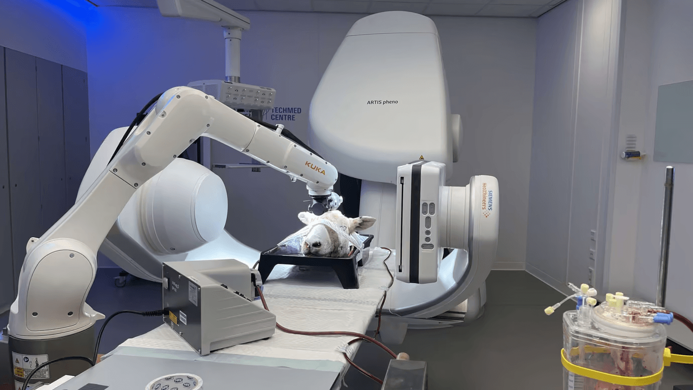
Researchers from the University of Twente (UT) and Radboud University Medical Center tested the device on sheep brain tissue in the lab, marking a step toward treating strokes, tumours, and other deep brain lesions without invasive surgery. The idea: a robot small enough to travel through the bloodstream, drill through an arterial wall at just the right spot, and continue into brain tissue to reach the problem directly.
How the magnetic robot works
After entering the body via the bloodstream, an external magnet spins the robot, which then screws itself forward, much like a corkscrew. Spin the magnet faster, and the robot speeds up too — but only up to a point. Past that threshold, known as the "step-out frequency," the robot stops keeping pace. It stalls, slips, or starts moving erratically.
Until now, that breaking point was largely guesswork. The team built a mathematical model, based on the Buckingham Pi theorem, that combines the robot's size, magnetic strength, and the tissue's stiffness to predict exactly when control is lost. Stiffer tissue lowers that threshold dramatically: in soft tissue, the robot kept up with the magnet at roughly 30 rotations per second, but in the stiffest tissue tested, that number dropped to less than one rotation per second.
"Push a magnetic robot too fast, and it simply stops listening to the magnet," said Ewout Ligtenberg, the study's first author. "We can now predict exactly when that happens, for any tissue, from a single test. That takes out a lot of guesswork when designing robots for the brain."
One test, endless predictions
Remarkably, the model only needs to be calibrated once, using a single measurement in tissue of known stiffness. From there, it can predict how the same robot design will behave in completely different tissue types, no extra testing required.
The team validated this first in gelatine, then in real sheep brain tissue. Without blood flow, the robot stayed in sync with the magnet up to about 1.8 rotations per second. But once blood was pumped through the vessels to mimic a living brain, that threshold dropped below 0.45 rotations per second — the added pressure made the tissue push back harder.
In practice, the robot drilled into brain tissue at a cautious 0.2 millimetres per second. Getting back out was much faster: retracing its own path, it moved at 2.9 millimetres per second, since it didn't need to carve a new channel. Using real-time camera tracking, the researchers also guided the robot to specific targets in a soft gel model of the brain, hitting them with sub-millimetre accuracy in some trials.
What's next
The work, published in Advanced Science, was carried out at UT's TechMed Centre and MESA+ institute, together with Michiel Warlé's team at Radboudumc. The robot's biocompatible coating was developed with Enschede-based LipoCoat B.V., with international input from the German University in Cairo, Triticum Medical, and the Hebrew University of Jerusalem.
There's still a long road from lab tissue to a human skull, but the milestone puts magnetically steered micro-robots a step closer to reaching the brain's hardest-to-treat corners without ever making a major incision.