Six top projects in Imaging Technologies to receive NWO funding
The HTSM program is closely aligned with the ten innovation domains of Holland High Tech and the National Technology Strategy (NTS).
Published on May 10, 2025

Team IO+ selects and features the most important news stories on innovation and technology, carefully curated by our editors.
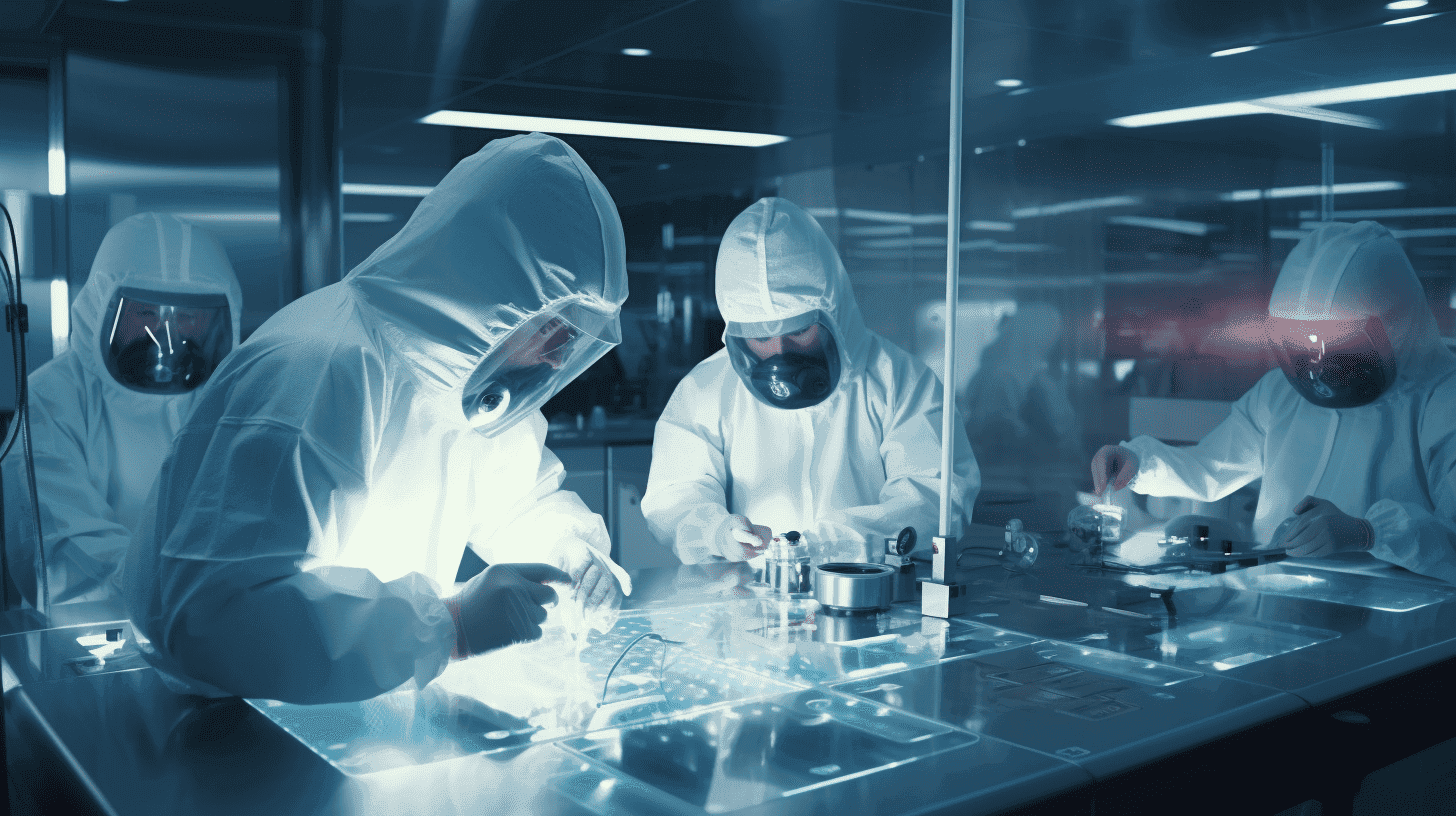
Six research projects into innovative techniques, including food quality testing, the development of less invasive MRI technology, the treatment of muscle diseases, and the development of molecular medicine for various types of cancer, will receive support from the NWO HTSM Call 2024. With this funding, the associated consortia, consisting of researchers from knowledge institutions and companies, will start research into Imaging Technologies, one of the ten priorities of the National Technology Strategy.
The high-tech sector plays a vital role in solving societal problems and creating economic opportunities for the Netherlands. Therefore, NWO and the High Tech Systems and Materials top sector (Holland High Tech) have joined forces to fund fundamental and application-oriented research. They are doing this through the HTSM program, which is aligned with the ten innovation domains of Holland High Tech and the National Technology Strategy (NTS). This strategy identifies key technologies needed for transitions such as the energy transition, the circular transition, and the development of digitization. Specifically, the research proposals that have now been awarded funding are linked to the key technologies Energy Materials and Imaging Technologies.
Leo Warmerdam, CEO of Holland High Tech, emphasizes the importance of Dutch high-tech companies' involvement in innovation: “This is precisely how knowledge and insights from research can quickly lead to value creation in the business community. The six projects will contribute to the development of the key technology Imaging Technologies, and thus also to our earning capacity in this important technology.”
NWO HTSM Call
The NTS identifies key technologies needed for urgent transitions, including the energy transition, the circular transition, and the development of digitization.
These are the projects that have been awarded funding:
1. DUAL-mode IMaging for Production and Automated Control Technologies (DUAL-IMPACT) - Leiden University
In manufacturing and maintenance industries, traditional quality control methods are often slow, labor-intensive, and error-prone. This is particularly problematic in sectors such as food processing and energy infrastructure, where rapid and accurate detection of contaminants or defects is essential to prevent risks to public health and safety. The need for faster, more reliable detection of internal defects is the motivation for this project. Advanced imaging techniques that combine detailed three-dimensional imaging with the efficiency and speed of high-throughput systems are required. The research within the project focuses on transforming industrial quality control by developing a new imaging workflow that integrates a '3D mode' into these systems.
Co-applicants: The Hague University of Applied Sciences (HHS), NWO institutes organization, CWI - Center for Science and Information Technology
Partners: Meyn Food Processing Technology, APPLUS
2. Cryogenic-soft landing imaging mass spectrometry - Maastricht University
Macromolecular assemblies (MMAs), such as protein complexes and viruses, play a crucial role in biological processes such as signal transduction and cellular transport. Understanding their molecular structure is essential for insights into human health. Cryogenic electron microscopy (cryo-EM) is a powerful tool for determining the atomic structure of MMAs. However, various challenges related to sample preparation, data collection and processing, electron beam damage, and instrumentation limitations hinder further progress in spatial resolution. The project focuses on image-guided native mass spectrometry (nMS) as a tool for sample preparation for cryo-EM.
Partners: Amsterdam Scientific Instruments, DEMCON, Thermo Fisher Scientific, VitroTEM
3. CLEAR-water: Contrast-agent Free Hemodynamic MRI - Leiden University Medical Center
Most MRI scans of the brain use a gadolinium-based contrast agent. The contrast agent leaves the patient's body via urine and ends up in sewage water. Gadolinium is difficult to filter out of surface water, and pollution levels above the limit values are often measured in Dutch river water. This makes it important to minimize the use of MRI contrast agents. This project is developing new MRI techniques that use magnetic labeling of the blood. This is a completely risk- and damage-free method that measures the same information without administering a contrast agent.
Partner: Philips
4. DeepSIM: Live tissue imaging at the nanoscale - TU Delft
Diseases such as cancer, where there are significant differences between patients, require treatments that are tailored to the individual. Molecular medicine can reduce healthcare costs by moving away from ineffective treatments. The necessary developments take time, and in an aging society, the urgency is high. A better understanding of the molecular origins of diseases is key to innovation, but this requires effective 3D imaging tools in the preclinical phase. The project aims to develop microscope technology and methodology that increases the spatial-temporal bandwidth, enabling faster, high-resolution 3D multi-label imaging in tissue.
Partners: Amsterdam Scientific Instruments, Confocal.nl, DELMIC, Lumicks, Flexible Optical (Okotech), Technolution
5. PRISTINE: PReclinical validation of multi-modal Imaging Systems and Technologies In NEuromuscular disorders - Radboud
Muscle diseases have a huge impact, not only on patients, but also on their families and close environment. These disorders limit daily functioning, lead to reduced quality of life, and cause both emotional and physical stress. This project aims to improve healthcare and the lifespan of individuals living with a muscle disease through the development of advanced multimodal imaging. A comprehensive preclinical muscle imaging tool is being developed. The multimodal imaging protocol will provide robust assessment of muscle contractility and tissue quality to characterize the changes in muscle health observed in the FLExDUX4 mouse model of facioscapulohumeral muscular dystrophy (FSHD).
Co-applicants: Leiden University Medical Center, University of Twente
Partners: iThera Medical, Solve FSHD
6. Advanced acquisition and image reconstruction for total-body positron emission tomography - University Medical Center Groningen
Positron emission tomography (PET), combined with computed tomography (CT), can be used to measure molecular interactions in both humans and animals. This capability significantly improves the diagnosis of many diseases, such as Alzheimer's and cancer. The application of PET imaging is limited by the amount of harmful ionizing radiation involved. To make PET imaging more widely applicable in medical practice, clinical research, and drug development, a significant reduction in radiation dose is essential. By significantly reducing radiation levels, this project will improve the safety of the technology for routine clinical use and expand its applications in both clinical settings and research.
Co-applicant: Amsterdam UMC
Partner: Siemens
