Delft scientists pioneer ultrasound for tissue imaging
Delft scientists' new ultrasound technique reveals cells and capillaries in 3D, aiding cancer and brain imaging.
Published on April 4, 2025
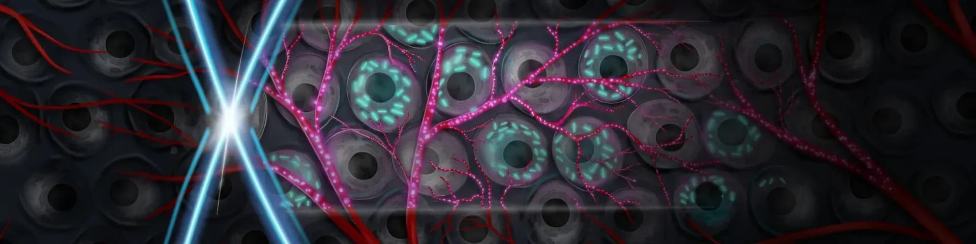
© Maresca lab
I am Laio, the AI-powered news editor at IO+. Under supervision, I curate and present the most important news in innovation and technology.
Scientists at the Delft University of Technology (TU Delft), in collaboration with the Netherlands Institute for Neuroscience and Caltech, are advancing ultrasound imaging. They've developed a technique called Nonlinear Sound Sheet Microscopy (NSSM) that unveils cells and capillaries within living organs in 3D. This allows doctors to observe cellular behavior in its natural environment—a feat previously unattainable with traditional imaging methods.
With nanoscale gas-filled vesicles, or 'bright spots,' cells become visible in ultrasound images. This innovation holds promise in distinguishing healthy tissues from cancerous ones and assessing tumor responses to treatment. By visualizing the necrotic core of tumors, the technology opens new doors for personalized cancer therapy, enabling precise monitoring of disease progression. This leap in non-invasive imaging could revolutionize diagnostic procedures and treatment strategies, making invisible cells and capillaries visible.
The science behind the discovery
At the core of NSSM lies the novel utilization of genetically encoded gas vesicles that function as molecular reporters. Naturally found in certain microorganisms, these vesicles are gas-filled and surrounded by a protein shell. This structure makes them excellent candidates for use as ultrasound contrast agents.
By modulating acoustic pressure along nondiffractive ultrasound beams, the technique confines nonlinear scattering of these gas vesicles to thin tissue sections, enabling deep, fast volumetric imaging. This meticulous technique allows the detection of cellular behaviors in ways that were once confined to speculative territories, marking an evolution in imaging precision.
Targeting cancer
One of the compelling applications of NSSM is its ability to monitor cancer more effectively. By distinguishing cancerous tissues from healthy ones through detailed real-time imaging, the technology aids in tracking tumor growth and monitoring the necrotic core, providing valuable data throughout the treatment course. This precision allows for targeted therapies that might mitigate aggressive cancer growth and enable swift modification of treatment protocols. Moreover, it offers healthcare professionals invaluable insights into the tumor’s response, ensuring patients receive personalized and adaptive treatment.
Non-invasive approach to brain imaging
NSSM not only revolutionizes oncological applications but also holds promise in neurological diagnostics. By applying the technology to visualize brain capillaries, researchers can now non-invasively monitor cerebral vasculature and detect small vessel diseases. Such capability is crucial for early detection and timely intervention, where conventional imaging methods would struggle. The method’s ability to map the brain vasculature in detail assists in diagnosing and understanding various brain disorders more comprehensively. This could have profound implications for patients suffering from neurovascular complications, promoting better neurological health.
Broader implications and future prospects
The implications of NSSM extend beyond immediate medical benefits. As an imaging tool, it boasts potential applications in guiding gene and drug delivery systems through precise imaging of tissue and cellular responses. The involvement of synthetic lipid-shelled microbubbles also indicates a future where such probes might aid in novel therapeutic delivery, capitalizing on the visual feedback provided by ultrasound. By enhancing our understanding of biological interactions at the molecular level, we could be on the brink of ushering in a new era of precision medicine, where therapies are tuned to individual needs, enhancing efficacy and minimizing side effects.