A new lens into the brain’s fiber maze
A light scattering technique reveals the complexity of the brain fiber structure paving the way for better diagnostic methods.
Published on November 26, 2025
© ScienceBrush

Mauro swapped Sardinia for Eindhoven and has been an IO+ editor for 3 years. As a GREEN+ expert, he covers the energy transition with data-driven stories.
Complexity is what makes the human brain truly special. A tight-knit network of neurons and cells that, despite operating on a minimal amount of power, achieves massive information computation capabilities. Much of the fascination we all have, scientists included, with the brain stems from its great mystery. An imaging technology called ComSLI has now proven to show in detail the orientation of brain fibers.
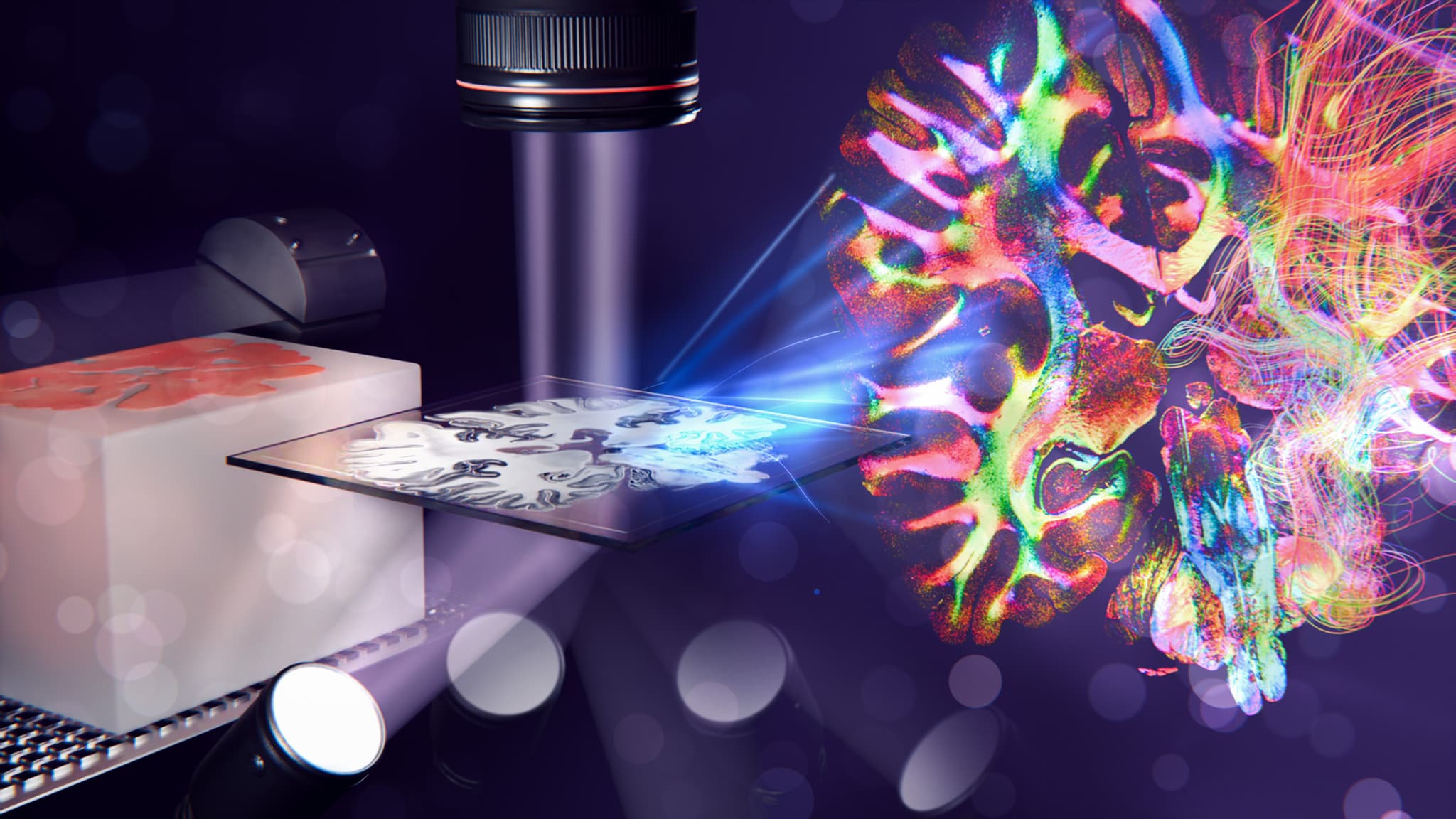
Miriam Menzel, an assistant professor at Delft University of Technology, has developed computational scattered light imaging (ComSLI). “The technique can disentangle the dense fiber network of brain tissues down to the micron level. Moreover, it can reveal the individual nerve fiber orientations, also in the regions with densely interwoven fibers, something that is not possible with other methods,” she explains.
Why are fiber orientations so important? Because they can, for example, study the progression of Alzheimer's or Parkinson's, providing scientists with better information on how these diseases unfold in the affected brain regions. Given the high density of the fiber network, disentangling the interwoven fibers has, until now, proven difficult.
In a new research paper, published alongside other scientists from TU Delft, Stanford University, Erasmus Medical Center of Rotterdam, and the German research center Forschungszentrum Jülich (FZJ), it was shown how this technique can be applied to a wide range of histological tissue sections. ComSLI can work regardless of the way the tissues are prepared. The paper was published in Nature Communications.
Lighting up the brain's fiber network
To study the brain’s anatomy under a microscope, brain tissue has to be cut into thin sections. The tissues used for this kind of research come from the brains of people who have passed away and are stored in clinical centers. To achieve high-quality, micrometer-thin sections, the tissue needs to be hardened in paraffin wax before sectioning. The preparation method is called formalin-fixed paraffin-embedding (FFPE). However, it comes with a significant downside: it compromises the nerve fibers’ optical properties, needed for other imaging techniques.
Conversely, ComSLI is not constrained by this limit. By illuminating a tissue sample from multiple angles and recording the transmitted light with a high-resolution camera, it is possible to capture how light scatters. "Light scatters mostly perpendicular to aligned structures like nerve or muscle fibers," says Menzel. Analyzing these scattering patterns for each micron-sized image pixel enables the researchers to compute local fiber orientations—even when multiple fibers cross within a single image pixel.
Moreover, unlike traditional methods, ComSLI doesn't require expensive lasers or complex setups. "You can build a basic version with a hobby camera and an LED ring in a day," Menzel notes. The resolution depends on the camera. Two to four microns are ideal for clinical applications.
The result is a color-coded map of all the fiber orientations. Each color represents a different angle, providing a detailed view of the fiber bundles connecting the different brain regions. In neurodegenerative diseases like Parkinson’s, the orientation of these fibers provides clues about disease progression and which areas are involved.
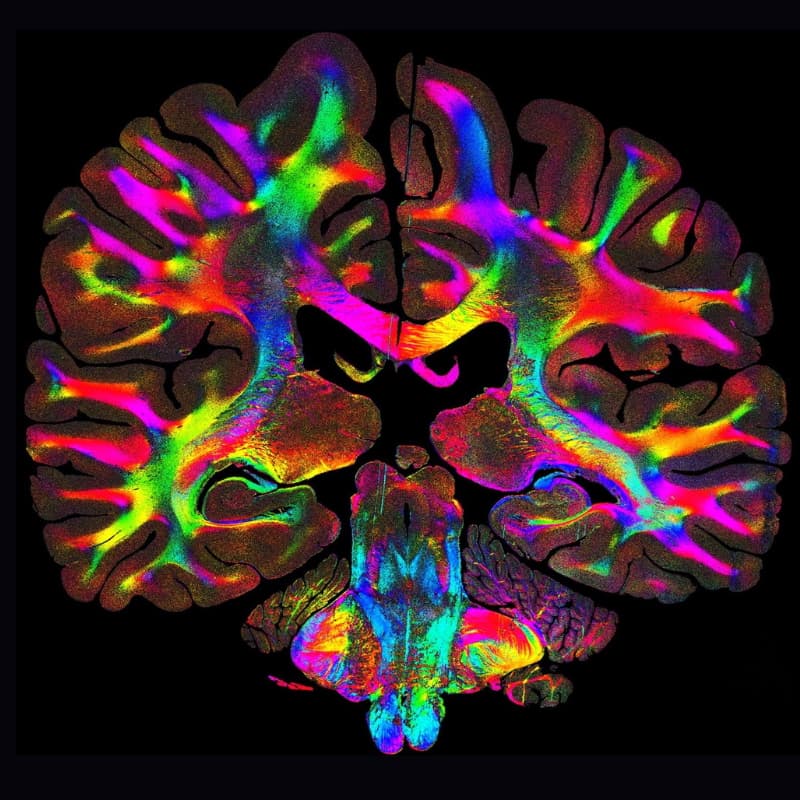
A scan of a brain section with the colored fibers. © Georgiadis et al., Nature Communications 16:9572, 2025
Applying physics to brain research
A physicist by training, Menzel, who has collected academic experience across Germany, the United Kingdom, and the United States, has found herself at the intersection of physics and neuroscience. "I was fascinated by the idea of using physics to address challenges in brain research," she recalls. Her early work focused on polarization microscopy, a technique that visualizes nerve fiber structures but struggles with densely packed or crossing fibers.
It was during her PhD at FZJ that she discovered that light scattering could reveal fiber orientations in a way polarization microscopy couldn't. This discovery paved the way to start developing the imaging method. Now, she heads a TU Delft lab entirely dedicated to ComSLI research.
"The brain is a never-ending story. There is always something new to learn about, by just tweaking the imaging parameters, new elements light up; it is very fascinating. Then, it is gratifying to see neuroscientists making sense of the images we can provide them and allow them to see things they have never seen before," Menzel emphasizes.

Dr. Miriam Menzel
Assistant professor at Delft Univerisity of Technology
After several academic experiences across Germany, the United Kingdom, and the United States, she joined the Department of Imaging Physics at Delft University of Technology in October 2022.
Many more potential applications
ComSLI can be used for many more applications—basically, wherever there is a fiber network. As shown by the researchers in the paper, the technique also reveals muscle and collagen fibers, as well as directed structures in bone and blood vessels. In addition, the method could also be potentially used for non-biological tissues. In materials science, investigating the fiber network provides a clearer picture of the material's structure.
Still, ComSLI has proven to be particularly apt for biomedical research. One promising direction is the study of collagen fibers in tumors. "Collagen organization at tumor boundaries can tell us more about the degree of cancer,” Menzel notes. “Our method can potentially visualize these changes, offering new insights for cancer diagnostics.”
The researcher's immediate goal is to make ComSLI more specific, making it able to distinguish between nerve, muscle, and collagen fibers in complex tissues. Bringing the technique into the clinics remains the primary goal to benefit patients directly.
In the long term, one of the most exciting prospects is intraoperative diagnostics—using the technique during surgeries to provide real-time tissue analysis. "Surgeons could use our method to assess tumor margins or brain fiber tracts quickly," Menzel envisions. "ComSLI is non-destructive, fast, and doesn't require staining. But there is a long way to go."